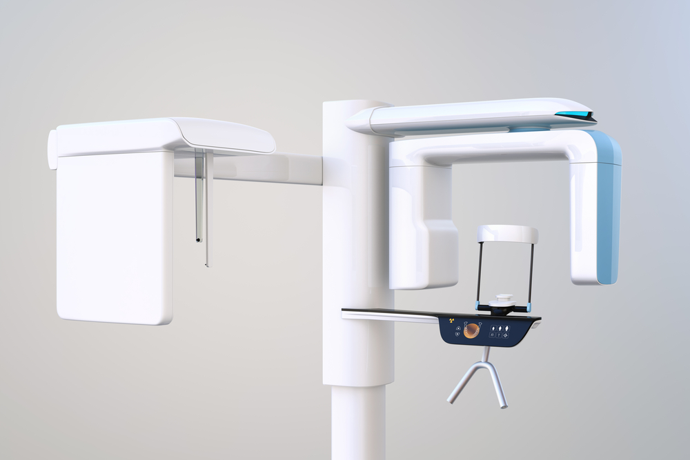
3D Cone Beam Computed Tomography (CBCT) is a revolutionary imaging technology that has dramatically improved the way dental professionals assess and diagnose conditions affecting the teeth, jaws, and surrounding structures. Unlike traditional two-dimensional X-rays, CBCT provides a highly detailed three-dimensional view, allowing for a deeper understanding of complex oral anatomy. This imaging system uses a cone-shaped X-ray beam that rotates around the patient to capture hundreds of images in under a minute, which are then compiled into a 3D model.
Because it offers a comprehensive view of hard tissues like bones and teeth, CBCT is particularly valuable in diagnosing conditions that may be missed by standard imaging. The level of detail enables dental professionals to plan treatments with greater precision, reducing the risk of complications and improving outcomes for patients. CBCT is now widely used across a variety of dental specialties, from endodontics and periodontics to implantology and orthodontics.
The Evolution of Dental Imaging Technologies
For decades, dental professionals relied on 2D X-rays such as bitewings, panoramic films, and periapical radiographs to guide diagnosis and treatment. While these tools remain useful, their limitations are well-known. Two-dimensional images flatten three-dimensional structures, which can result in overlaps and blind spots, making it difficult to locate certain abnormalities or understand anatomical relationships.
The development of CBCT marked a major milestone in dental imaging. First introduced in the early 2000s, it quickly gained popularity due to its ability to generate 3D images with relatively low radiation doses compared to conventional CT scans. The technology filled a crucial gap between traditional dental radiographs and the high-radiation, hospital-based medical CT systems, offering dentists a powerful yet accessible diagnostic tool.
When and Why CBCT is Used in Dentistry
CBCT is typically not used for routine dental check-ups but is reserved for more complex cases where additional anatomical detail is essential. Dentists may recommend CBCT scans for evaluating impacted teeth, assessing bone structure before placing dental implants, or identifying root fractures that cannot be seen with traditional X-rays.
This technology has become invaluable for oral surgeons, periodontists, and endodontists who need to visualize the precise position of nerves, sinuses, and root canals before performing delicate procedures. CBCT is also a go-to imaging tool in orthodontics for evaluating jaw relationships, airway structures, and craniofacial development.
Key Benefits of Cone Beam Imaging
- Improved Diagnostic Accuracy: CBCT offers detailed visualization of bone, teeth, and surrounding tissues from multiple angles. This makes it easier to detect conditions like cysts, abscesses, and root fractures that may not appear clearly on 2D X-rays.
- Enhanced Treatment Planning: The 3D data allows for accurate mapping of dental implants, assessment of jawbone density, and evaluation of proximity to critical anatomical structures, reducing surgical risks.
- Minimally Invasive: Because CBCT enables detailed planning, many surgical procedures become less invasive. Surgeons can anticipate complications and avoid unnecessary tissue trauma.
- Lower Radiation than Conventional CT: Compared to medical-grade CT scanners, CBCT uses a much lower radiation dose, making it a safer option for dental-specific applications.
- Faster and More Comfortable for Patients: The scanning process takes under a minute and requires no uncomfortable bitewings or positioning. Patients stand or sit still while the machine rotates around the head.
Comparing CBCT to Traditional Imaging
While traditional dental X-rays are useful for general exams and monitoring oral health over time, they fall short when precise details are required. For example, a 2D panoramic image might show an impacted wisdom tooth but won’t reveal the tooth’s proximity to the inferior alveolar nerve—a critical detail that CBCT captures.
Another comparison is in endodontics. Locating complex root canal systems with 2D images can be challenging, especially in molars. CBCT removes the guesswork, allowing the dentist to see the exact shape and orientation of the canals before treatment. This leads to greater success rates and reduced need for retreatment.
Common Applications Across Dental Specialties
CBCT’s versatility extends into nearly every branch of dentistry. Some of the most common uses include:
- Dental Implant Planning: Assessing bone volume, density, and orientation of neighboring structures
- Orthodontics: Evaluating jaw alignment, impacted teeth, and airway dimensions
- Oral Surgery: Locating nerves, roots, and sinuses to minimize surgical risks
- Endodontics: Identifying hidden canals, root fractures, and periapical lesions
- TMJ Analysis: Visualizing temporomandibular joint anatomy for diagnosis of disorders
These applications underscore the importance of CBCT as not just a diagnostic tool, but as a roadmap for personalized, high-precision treatment planning.
What Patients Can Expect During a CBCT Scan
Many patients are unfamiliar with the CBCT process and may feel anxious about the scan. In reality, the procedure is quick, painless, and requires minimal preparation. Patients are usually asked to remove metal items like earrings or glasses before the scan. They then either sit or stand in the CBCT unit while the machine performs a full rotation around their head, typically in under 40 seconds.
There’s no need for injections, contrast dyes, or sedation. Once the scan is complete, the digital images are immediately available for review. Dentists can manipulate the 3D model on a computer screen to explore different angles, zoom in on specific areas, or create cross-sectional slices for analysis.
Safety Considerations and Radiation Exposure
Understandably, patients often ask about radiation exposure. While CBCT does involve more radiation than a single periapical X-ray, it’s significantly lower than that of a conventional CT scan. The effective dose can vary depending on the size of the scan area and machine settings, but modern CBCT units are designed with dose-reduction protocols to prioritize patient safety.
Dentists weigh the risks and benefits before recommending a CBCT scan. When the clinical need is justified, the diagnostic value outweighs the minimal radiation exposure. Moreover, because CBCT often prevents the need for exploratory procedures or additional scans, it can actually reduce overall exposure in the long term.
Limitations and Considerations
Despite its many advantages, CBCT is not without limitations. The technology excels at capturing hard tissue structures but is less effective at imaging soft tissues like gums or lymph nodes. It also requires interpretation by trained professionals, and the vast amount of data can sometimes reveal incidental findings that may not be clinically relevant.
Additionally, not all dental offices are equipped with CBCT machines due to the cost and space requirements. In some cases, patients may be referred to a radiology center or specialty clinic to obtain the scan. This underscores the importance of patient education—understanding why the scan is being recommended can help alleviate concerns about convenience or cost.
The Future of CBCT in Dental Care
As imaging technology continues to evolve, CBCT is expected to play an even more prominent role in modern dentistry. Advances in software now allow dentists to integrate CBCT data with CAD/CAM systems for designing surgical guides, aligners, and restorations with unprecedented precision.
Artificial intelligence is also beginning to shape how CBCT images are analyzed. Automated algorithms can help detect abnormalities, measure structures, and generate treatment recommendations, streamlining the diagnostic process while reducing the risk of human error.
For patients, this means faster diagnosis, more predictable results, and an overall improved dental experience. As awareness grows, CBCT is likely to become the gold standard for complex dental evaluations.
Final Thoughts on CBCT’s Role in Modern Dentistry
3D Cone Beam Imaging represents a major leap forward in dental diagnostics and treatment planning. By offering a comprehensive, three-dimensional view of oral anatomy, CBCT has changed the way many dental procedures are performed—making them safer, more effective, and more tailored to each patient. While not necessary for every dental visit, it remains a powerful tool for complex cases that require precision and clarity beyond what traditional X-rays can provide.
Whether you’re preparing for a dental implant, orthodontic evaluation, or endodontic treatment, understanding the role of CBCT can help you make informed decisions about your care. As with all aspects of dental health, communication with your provider is key. Don’t hesitate to ask questions and explore how imaging technologies may support your treatment plan.
Resources
Patel, S., Horner, K., & Whaites, E. (2015). Cone Beam CT in Dental Practice. British Dental Journal.
Scarfe, W. C., & Farman, A. G. (2008). What is Cone-Beam CT and How Does it Work? Dental Clinics of North America.
Al-Rawi, B., Hassan, B. A., & Vandenberge, B. (2010). Cone Beam Computed Tomography (CBCT) Imaging of the Craniofacial Region: A Review. International Journal of Dentistry.